
Diagnostic Ultrasound
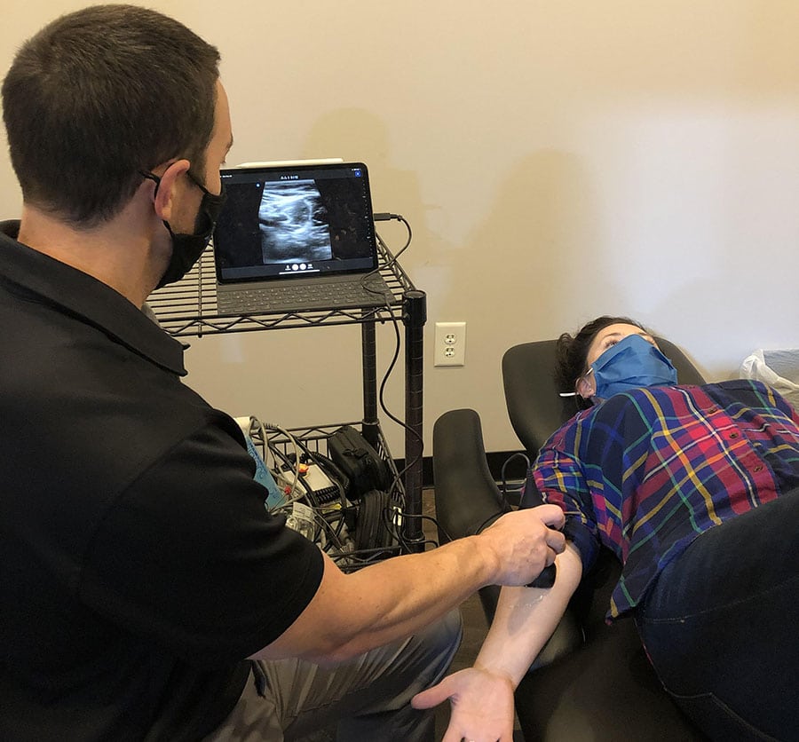
Ultrasound imaging uses high-frequency sound waves to allow viewing of structures beneath the skin’s surface. The image is produced by the reflection of the waves off the body structures being examined. The image is produced instantly and allows the provider to immediately assess the status of the tissues.
Procedure
First, a thin layer of ultrasound gel is applied to the skin. This allows the sound waves to easily pass from the transducer head into the body and back to create an image. There is no pain associated with the procedure and the patient does not feel anything more than the ultrasound head being moved over the skin’s surface. The procedure itself usually only takes a few minutes.
Uses
There are a wide variety of uses for diagnostic ultrasound. Many people may be familiar with ultrasound used to visualize a fetus within a mother’s womb. In some settings, it can be used to assess blood flow through a vessel or organ. In the orthopedic world and at PRA, we are using ultrasound to assess tissue quality. It can help diagnose muscle and tender tears, calcifications, scar tissue, or abnormal growths.
Diagnostic ultrasound allows the provider to assess soft tissues whereas an x-ray would mainly show bony structures. There is also no radiation exposure with ultrasound use which makes it safer than an X-ray. MRIs will give a deeper, more detailed view of soft tissues but this is not always necessary for a proper diagnosis. An MRI is also very expensive, and ultrasound can be an adequate, more cost-effective option. We are excited to now offer this diagnostic procedure here at PRA! Your provider can make a recommendation if ultrasound imaging may be necessary for proper diagnosis and direct your treatment in the right direction to get you better, and faster.
Sources
https://www.osc-ortho.com/blog/diagnostic-ultrasound-for-orthopaedics/
https://www.fda.gov/radiation-emitting-products/medical-imaging/ultrasound-imaging

All Services
Learn More
Keep
In Touch
Cotswold
(980) 819-8545

Office Hours
| Monday: | 7:00 am–6:00 pm |
| Tuesday: | 8:20 am–6:00 pm |
| Wednesday: | 7:00 am–4:30 pm |
| Thursday: | 8:40 am–6:00 pm |
| Friday: | 7:00 am–3:00 pm |
| Saturday: | Closed |
| Sunday: | Closed |
Lake Norman
Office Hours
| Monday: | 8:00 am–6:00 pm |
| Tuesday: | Closed |
| Wednesday: | 7:00 am–6:00 pm |
| Thursday: | 8:00 am–4:00 pm |
| Friday: | 7:00 am–12:00 pm |
| Saturday: | Closed |
| Sunday: | Closed |
South Charlotte
Office Hours
| Monday: | 7:00 am–6:00 pm |
| Tuesday: | 7:00 am–6:00 pm |
| Wednesday: | 7:00 am–6:00 pm |
| Thursday: | 7:00 am–6:00 pm |
| Friday: | 7:00am–3:00pm |
| Saturday: | Closed |
| Sunday: | Closed |
© 2024 Performance Rehab Associates. All Rights Reserved. Accessibility Statement - Privacy Policy - Sitemap


